
Reviewed by Dr. Venkatesh Mishra (BPT, MPT, Certified Dry Needling Practitioner) — VS Physiotherapy, Osteopathy & Chiropractic Clinic
Most people meet the phrase “L4-L5 disc bulge” for the first time on an MRI report, in a paragraph they cannot read, handed to them by someone with three minutes to explain it. By the time they get home and start searching, two things have usually happened: the language has started to sound frightening, and someone has mentioned surgery.
This guide is written to undo both of those. It explains what the L4-L5 segment is, what each phrase in your MRI report actually means, how to tell which nerve root is involved from your own symptoms, what genuinely helps, and the narrow set of circumstances in which surgery is the right answer.
Why L4-L5 Fails More Often Than Any Other Level
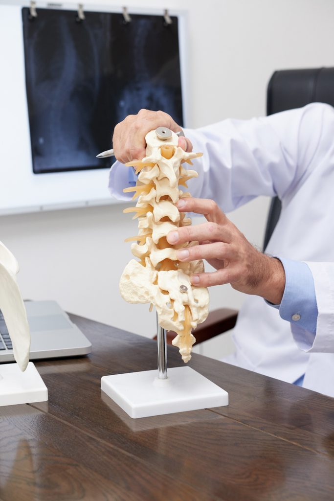
Your lumbar spine has five vertebrae, stacked L1 through L5, sitting on the sacrum. Between each pair is a disc, a tough fibrous ring (the annulus fibrosus) wrapped around a soft gel core (the nucleus pulposus).
L4-L5 sits second from the bottom, and it works harder than almost anything else in the body:
- It carries the most load. Everything above it — head, arms, trunk is transmitted through this segment every time you stand, lift, or lean forward.
- It moves the most. L4-L5 contributes the largest share of flexion and extension in the lumbar spine. Load plus movement is the combination that wears tissue out.
- It sits at a transition. Below it, L5-S1 hands off to the sacrum, which is fixed. Segments next to rigid structures take more shear stress.
This is why L4-L5 and L5-S1 account for the overwhelming majority of lumbar disc problems, and why L4-L5 is also the most common site of degenerative spondylolisthesis.
Bulge, Protrusion, Extrusion, Sequestration: They Are Not the Same Thing
Radiologists use these four words precisely. Patients and unfortunately many websites use them interchangeably. The difference matters, because it changes both the likely symptoms and the likely course.
| Term | What it means | How serious |
| Disc bulge | The disc extends beyond the vertebral rim around a wide arc, more than 25% of its circumference. The annulus is still intact. | Usually the mildest. Very common with age. Often causes no symptoms at all. |
| Protrusion | A focal outpouching over less than 25% of the circumference. The base of the outpouching is wider than its tip. | Moderate. More likely to contact a nerve root because it is focal. |
| Extrusion | Nucleus material has pushed through the annulus. The tip is now wider than the base, it has squeezed out like toothpaste. | More significant. Also, paradoxically, more likely to shrink on its own. |
| Sequestration | A fragment of disc material has broken free and is sitting separately in the spinal canal. | Sounds worst. Has the highest rate of spontaneous resorption of all four. |
Hold on to that last column. It contradicts what almost everyone assumes, and we will come back to it.
How to Read Your Own MRI Report
This is the section most people actually came for. Here is what the standard phrases mean, in plain language, with an honest note on whether each one should worry you.
“Diffuse disc bulge at L4-L5” The disc is bulging fairly evenly around a broad arc rather than in one focal spot. This is the mildest and most common finding on lumbar MRI. On its own, it is a description of a disc that has aged, not a diagnosis of the cause of your pain.
“Posterocentral” / “paracentral” / “foraminal” / “far-lateral” These describe where the bulge points, and they are the single most useful words in your report.
- Posterocentral — straight backwards, towards the middle of the canal.
- Paracentral (posterolateral) — backwards and slightly off to one side. This is the most common direction, because the posterior longitudinal ligament is weakest here.
- Foraminal / far-lateral — out to the side, into or beyond the tunnel where the nerve root exits.
Direction determines which nerve is affected. We will map that out in the next section.
“Indentation of the thecal sac” / “effacement of the anterior thecal sac” The thecal sac is the fluid-filled membrane containing the nerve roots. Mild indentation means the disc is touching it. It is an extremely common finding and does not by itself mean a nerve is being compressed or that you need surgery.
“Neural foraminal narrowing” (mild / moderate / severe) The exit tunnel for the nerve root is narrower than normal. The grading matters far more than the phrase. Mild narrowing is frequently symptom-free; severe narrowing correlates better with radiating leg symptoms.
“Annular tear” or “high-intensity zone (HIZ)” A fissure in the outer fibrous ring, showing up as a bright spot on T2 images. Annular tears can be a source of localised back pain, and they are also found in people with no pain at all.
“Disc desiccation” / “loss of T2 signal” The disc has lost water content and appears dark. This is a normal ageing change. Nearly every adult over forty has some of it.
“Modic changes” (Type I, II, or III) Signal changes in the bone marrow of the vertebral endplates next to the disc. Type I suggests active inflammation and oedema and can be associated with more pain; Type II is fatty replacement and is generally more stable; Type III is sclerosis.
“Ligamentum flavum hypertrophy” / “facet arthropathy” The ligament at the back of the canal has thickened, or the small facet joints have developed arthritic change. These often contribute more to canal narrowing than the disc itself does, particularly in older patients.
The most important thing to understand about your MRI
Large studies of people with no back pain whatsoever have scanned them anyway. The findings are consistent and worth sitting with: disc bulges are present in roughly 30% of pain-free 20-year-olds and in the substantial majority of pain-free 70- and 80-year-olds. Disc degeneration follows the same curve, appearing in around 90% of pain-free people by their sixties.
Your MRI describes the structure of your spine. It does not, on its own, explain your pain. A bulge that matches your symptom pattern is meaningful. A bulge that matches nothing you feel is an incidental finding and treating incidental findings aggressively is how people end up with unnecessary procedures.
This is why a proper clinical examination matters more than the report. The scan is one input.
Which Nerve Is It? L5 Root vs L4 Root
At L4-L5, two different nerve roots can be affected, and which one depends on the direction of the bulge. The symptom patterns are distinct enough that you can often work it out yourself.
The L5 root — compressed by a paracentral or posterocentral bulge
This is by far the more common of the two, because most bulges point backwards and slightly to one side, where they catch the L5 root as it travels down past the segment.
- Pain path: buttock → outer (lateral) thigh → outer calf → across the top of the foot → into the big toe.
- Numbness or tingling: top of the foot and the web space between the first and second toes.
- Weakness: difficulty lifting the front of the foot (ankle dorsiflexion) and difficulty raising the big toe against resistance. Severe cases produce a foot drop — the front of the foot slaps down when you walk, or you catch your toe on steps. Hip abductor weakness can also appear, causing a hip drop when standing on one leg.
- Reflexes: usually unchanged. There is no reliable, easily tested reflex for L5, which is precisely why L5 problems are sometimes missed.
- Provocation: typically worse with sitting, bending forward, coughing, or sneezing. A straight leg raise often reproduces the leg symptoms.
The L4 root — compressed by a foraminal or far-lateral bulge
Less common, but frequently misdiagnosed as a hip or knee problem.
- Pain path: front and outer thigh → across the knee → down the inner (medial) shin.
- Numbness or tingling: inner side of the lower leg and around the medial knee.
- Weakness: the quadriceps. Knee gives way on stairs, difficulty rising from a low chair, difficulty straightening the knee against resistance.
- Reflexes: the knee-jerk (patellar) reflex is reduced or absent on the affected side. This is a genuinely useful diagnostic sign.
- Provocation: often worse with extension and walking rather than sitting. The femoral nerve stretch test, bending the knee while lying face down, reproduces front-of-thigh symptoms
If your symptoms sit clearly in one of these two columns, and your MRI describes a bulge in the corresponding direction, the picture is coherent and treatment can be targeted precisely. If they do not match, the disc may not be the whole story and that is worth knowing before anyone operates.
Red Flags: Go to a Hospital Today
Most L4-L5 disc problems are not emergencies. A small number are. Do not wait for an appointment if you develop:
- Difficulty passing urine, loss of bladder control, or loss of bowel control
- Numbness in the saddle area — inner thighs, buttocks, genital region, the parts that would contact a saddle
- Weakness or numbness in both legs at once
- New sexual dysfunction alongside back and leg symptoms
- Rapidly progressing weakness — a foot drop that is clearly worse this week than last
Together these can indicate cauda equina syndrome, compression of the bundle of nerve roots at the base of the spinal cord. It is a surgical emergency, and outcomes depend heavily on how quickly it is decompressed. Go directly to a hospital emergency department, not to a physiotherapy clinic.
Also seek prompt medical review for back pain accompanied by unexplained weight loss, fever, night pain that wakes you consistently, or a history of cancer.
Does an L4-L5 Disc Bulge Heal on Its Own?
The honest answer has two parts, and most articles only give you the flattering half.
Part one: disc material genuinely does resorb. The body treats extruded nucleus material as something foreign, and immune cells break it down. Pooled research shows spontaneous regression occurring in roughly two-thirds of lumbar disc herniations overall and the rate is highest for the types that sound most alarming. Sequestrated fragments resorb most often. Extrusions resorb frequently. The general principle is that the further the material has travelled outside the disc, the more the body clears it.
Part two: a simple bulge is the least likely of all four types to disappear. Because the annulus is intact and nothing has escaped into the canal, there is little for the immune system to clear away. A diffuse bulge at L4-L5 will often still be visible on a scan years later.
That sounds like bad news. It is not, and here is why it matters more than anything else in this article:
Symptom recovery does not require the bulge to disappear. People get better when the inflammation around the nerve root settles, when the nerve stops being mechanically irritated with every movement, and when the surrounding muscles start sharing load properly again. All three of those can happen while the bulge remains exactly where it is. Plenty of people are completely pain-free with a bulge still sitting on their scan, remember the asymptomatic-population data above.
So the goal of treatment is not to chase a picture. It is to calm the nerve, restore movement, and rebuild capacity.
Realistic timelines with proper conservative care:
- Mild cases: meaningful relief in 4-6 weeks
- Moderate cases with radiating leg symptoms: substantial improvement over 8-12 weeks
- Severe or long-standing cases: gradual, steady progress over 12-16 weeks
Anyone promising to fix an L4-L5 disc bulge in a week is selling something.
How Non-Surgical Treatment Actually Works
Understanding the mechanism helps you tell useful treatment from expensive theatre.
A disc under load is a pressurised structure. Sitting raises pressure inside the lumbar discs considerably compared with standing or lying, and forward bending raises it further. That sustained pressure does two things: it pushes disc material outward against the annulus and nerve root, and it prevents the disc from doing the one thing it needs to repair itself.
Discs have almost no direct blood supply. They receive nutrients through diffusion across the vertebral endplates, and that diffusion works best when pressure inside the disc is low which is why you are measurably taller in the morning than at night. A disc under constant compression is a disc that cannot feed itself.
Mechanical unloading targets exactly this. Spinal decompression therapy uses a motorised table that applies graded, computer-controlled axial traction in a cycle of pull and release, rather than the constant static pull of older traction machines. The cycling matters: it avoids triggering the protective muscle guarding that a sustained pull provokes, which is the main reason simple traction historically underperformed.
The intended effects are a reduction in pressure inside the targeted disc, improved fluid and nutrient exchange across the endplates, and reduced mechanical irritation of the nerve root, giving inflammation a chance to settle.
An honest note on the evidence: the research base for motorised decompression is promising but not conclusive, and it is stronger as part of a structured rehabilitation programme than as a standalone cure. Any clinic presenting it as a guaranteed fix in isolation is overselling it. Used correctly as the unloading component of a phased plan, it does useful work.
The Treatment Sequence That Works
Effective care for an L4-L5 bulge is sequenced. Skipping ahead is the most common reason people plateau.
Phase 1 — Calm the nerve (roughly weeks 1-3)
The priority is reducing inflammation around the nerve root and breaking the pain-spasm cycle. Nothing else progresses until this happens.
- Class-4 laser therapy — photobiomodulation delivered deep enough to reach lumbar structures, used to reduce perineural inflammation and support tissue repair.
- Super Inductive System (SIS) — high-intensity electromagnetic field therapy for pain modulation and relief of the deep paraspinal spasm that accompanies acute disc irritation.
- TENS and IFT — electrical pain modulation, useful for making the early days tolerable enough to keep moving.
- Activity modification — not bed rest. Prolonged rest reliably makes disc problems worse. Short, frequent walks are one of the most effective things you can do in week one.
Phase 2 — Unload the segment (roughly weeks 2-8, overlapping)
Once the acute flare has settled enough to lie comfortably, mechanical unloading begins.
- Spinal decompression — a course of sessions on the decompression table, with the angle of pull adjusted to target L4-L5 specifically and force progressed gradually across the course. Sessions run alongside rehabilitation, not instead of it.
- Manual therapy and mobilisation — graded joint mobilisation to restore segmental movement without provoking the nerve root.
- Dry needling — the paraspinals, quadratus lumborum, gluteus medius and piriformis almost always develop secondary trigger points that outlast the original problem and produce their own referred pain. Treating them is often what breaks a stubborn plateau.
Phase 3 — Rebuild capacity (weeks 6-16)
This is the phase that determines whether you are back here in eighteen months.
- Motor control retraining — deep abdominal and multifidus activation, then integration into movement.
- Pilates mat work — low-load, high-control training that suits a recovering lumbar spine well.
- Progressive back care programming — graded loading until the spine tolerates the demands of your actual life, whether that is a desk, a shop counter, or a two-wheeler commute.
- Ergonomic and lifting correction — the piece almost everyone skips and almost everyone needs.
Where Chiropractic Fits, and Where It Does Not
This deserves a straight answer, because there is a great deal of noise around it.
Spinal manipulation — the high-velocity, low-amplitude adjustment has a reasonable evidence base for non-specific low back pain and for restoring movement at stiff segments. In a lumbar spine that has become globally guarded around an irritable disc, restoring segmental motion can meaningfully reduce pain.
But it is not the first tool for every disc presentation, and it should not be. Where there is acute, highly irritable radiculopathy, particularly with any measurable neurological deficit — gentler joint mobilisation, mechanical unloading and inflammation control come first. Manipulation, if it is used at all, comes later, once the nerve has settled.
Manipulation of the lumbar spine should be avoided outright where there is:
- Any suspicion of cauda equina syndrome
- Progressive motor weakness, such as a worsening foot drop
- Severe, unremitting radicular pain with clear neurological signs
- Significant osteoporosis, inflammatory arthropathy, suspected fracture, or malignancy
The right framing is straightforward: adjustment is one option within a plan, selected after examination, not a default applied to everyone who walks in with a scan. Any practitioner — of any discipline who proposes the same treatment before examining you is worth leaving.
Exercises by Phase
Two rules govern everything below.
The centralisation rule. If an exercise pulls your symptoms up the leg towards your back — calf pain becoming thigh pain, thigh pain becoming buttock pain — that is centralisation and it is a good sign, even if the back pain briefly increases. If symptoms travel further down the leg, that is peripheralisation. Stop that exercise.
The extension-preference rule. Many but not all L4-L5 bulges respond well to backward bending. Test it before you commit to it.
| Phase | Exercise | Dosage |
| Acute (weeks 1-3) | Prone lying, progressing to prone on elbows | 5-10 min, 2-3× daily |
| Prone press-up — only if it centralises | 10 reps, every 2-3 hours | |
| Supine pelvic tilt | 10 reps × 5 sec hold, 2× daily | |
| Sciatic nerve slider (glide, not stretch) | 10 reps, 3× daily | |
| Short walks | 5-10 min, 4-6× daily | |
| Subacute (weeks 3-8) | Dead bug | 8-10 per side × 2-3 sets |
| Bird dog | 8-10 per side × 2-3 sets | |
| Glute bridge | 10-15 reps × 3 sets | |
| Side plank from knees | 15-20 sec × 3 per side | |
| Hip flexor stretch | 30 sec × 2 per side | |
| Return to load (weeks 8+) | Hip hinge pattern, unloaded then loaded | 10-12 reps × 3 sets |
| Suitcase carry | 20-30 m × 3 per side | |
| Split squat | 8-10 per side × 3 sets |
Nerve glides are worth a specific note. They are sliders, not stretches — the nerve is mobilised gently through its path, never pulled taut. Aggressive hamstring stretching with an irritated L5 root reliably makes things worse, and it is one of the most common self-inflicted setbacks we see.
What to Avoid
- Loaded lumbar flexion. Sit-ups, crunches, toe-touch stretches, and bent-back lifting all spike intradiscal pressure in exactly the wrong direction. Remove them entirely in the early phases.
- The first hour after waking. Discs are maximally hydrated overnight, so pressure and stiffness are highest first thing. This is the worst possible time to bend forward, touch your toes, or lift something heavy.
- Prolonged sitting. Sitting loads lumbar discs more than standing. Cap it at 20-30 minutes before standing and moving, even briefly.
- Lifting combined with twisting. The single most reliable way to re-injure a healing disc.
- Long two-wheeler rides on rough roads. Repetitive vertical vibration is genuinely hard on a recovering lumbar disc. If unavoidable, use a lumbar cushion and break the journey.
- Complete rest. More than a day or two of lying down measurably slows recovery.
The back belt question
Lumbar support belts have a narrow, legitimate role: short-term use during an acute flare, or during a specific unavoidable heavy task. Used that way they can reduce pain and let you keep moving.
Worn habitually, they work against you. The abdominal and spinal muscles that should be stabilising your spine begin to offload the work onto the belt and decondition. Evidence for belts as a long-term preventive measure is weak. Practical rule: no more than 2-3 hours a day, only during flares or heavy loading, and never as a substitute for building core strength.
Sleeping and Sitting Positions
Sleeping. The goal is keeping the lumbar spine near neutral so the nerve root is not compressed for eight hours.
- Side-lying: knees slightly bent with a firm pillow between them. This stops the top leg dropping forward and rotating your pelvis. A small towel roll under the waist helps if you have a defined waist and the mattress lets your side sag.
- On your back: a pillow under the knees to reduce lumbar extension and relax the hip flexors.
- Face down: generally the least suitable, since it extends the lower back while forcing the neck into rotation. If you have a strong extension preference and genuinely find it comfortable, a thin pillow under the abdomen makes it more tolerable.
- Mattress: medium-firm outperforms very firm in the available research. A rock-hard surface is not the virtue Indian households often assume it is.
- Getting out of bed: log-roll onto your side, drop the legs off the edge, and push up with your arms. Sitting straight up from lying is a flexion movement with poor leverage.
Why nights can be worse. Lying still for hours means no movement to disperse inflammatory mediators, and discs rehydrate and swell overnight, increasing pressure. Both effects are normal and both improve as the condition settles.
Sitting. This is where most L4-L5 patients do the real damage, one workday at a time.
- Preserve a small inward curve in the low back — a lumbar roll or rolled towel at belt height does this better than most ergonomic chairs.
- Hips slightly higher than knees, feet flat on the floor.
- Screen at eye level so you are not craning forward, which drags the whole spine into flexion.
- Stand and move every 30 minutes. This matters more than the chair.
- Avoid low, soft sofas and prolonged cross-legged floor sitting during the acute phase — both force sustained lumbar flexion.
When Surgery Is Genuinely Indicated
The great majority of L4-L5 disc bulges never need an operation. Surgery is clearly indicated in a narrow set of situations:
- Cauda equina syndrome — an emergency, as above.
- Progressive or severe motor weakness — a foot drop that is deteriorating, or quadriceps weakness that is worsening despite proper care.
- Disabling radicular pain that has not responded to genuinely adequate conservative treatment over roughly 6-12 weeks. “Adequate” means a structured programme actually completed, not painkillers and a leaflet.
- Recurrent, disabling episodes that repeatedly interrupt work and daily life.
Worth knowing when you weigh the decision: comparative studies of surgical versus conservative management of lumbar disc herniation consistently show that surgery produces faster relief, while outcomes between the two groups converge over the following years. Surgery buys speed. For most people it does not buy a better final destination — which is precisely why an unhurried, properly executed conservative trial is reasonable when there is no red flag and no progressive deficit.
Which Specialist Should You Consult?
- Physiotherapist — appropriate first contact for most cases. Clinical assessment, nerve root testing, and the rehabilitation programme that does the actual recovery work.
- Chiropractor or osteopath — manual care, joint mobilisation, and adjustment where indicated, best delivered within a broader rehabilitation plan rather than as isolated sessions.
- Orthopaedic spine surgeon or neurosurgeon — required if there are red flags, progressive neurological deficit, or failure of a genuine conservative trial. Also the right person for any surgical decision.
- Pain physician — for epidural steroid injection when pain is severe enough to prevent participation in rehabilitation. An injection is a window of opportunity to do the rehab, not a treatment in itself.
- Rheumatologist — if the pattern suggests inflammatory back pain rather than mechanical: prolonged morning stiffness, night pain that improves with movement, onset before age 45.
In practice, a clinic that can assess you clinically, deliver the unloading and pain-control work, and run the rehabilitation under one roof removes a lot of friction from the process — particularly across a 12-week programme where the phases need to hand off to each other cleanly.
When to Get Assessed
If you have leg symptoms that follow one of the nerve root patterns above, if back pain has persisted beyond two weeks without improving, or if you are holding an MRI report you cannot interpret, a proper clinical examination is the sensible next step. Not because the report is necessarily bad news — usually it is not — but because the examination is what tells you whether the finding on the scan is the thing causing your pain.
At VS Physiotherapy, Osteopathy & Chiropractic Clinic in Lucknow, Dr. Venkatesh Mishra and the team assess lumbar disc presentations with full neurological testing, then build the phased programme around what the examination actually shows — combining non-surgical spinal decompression, pain-modulation modalities, manual therapy and progressive rehabilitation.
You can read more about our approach to slip disc treatment and sciatica treatment, or about telling a disc problem apart from a simple muscle strain.
Call or WhatsApp: +91 7007932170
Clinic: C-18, Kalyanpur (West), Front of SRM (TATA) Motors, Mazar Waali Gali, Tedhi Pulia Ring Road, Lucknow, Uttar Pradesh 226022
This article is for general information and does not replace individual clinical assessment. If you have any of the red-flag symptoms described above, seek emergency medical care immediately.